Logo

Bone Mineral Density (BMD) & Body Composition Scan

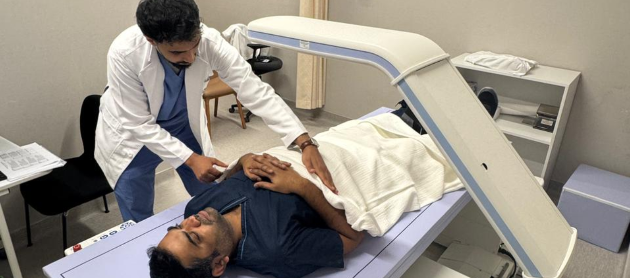
Insight that protects your strength and body.

contact-img

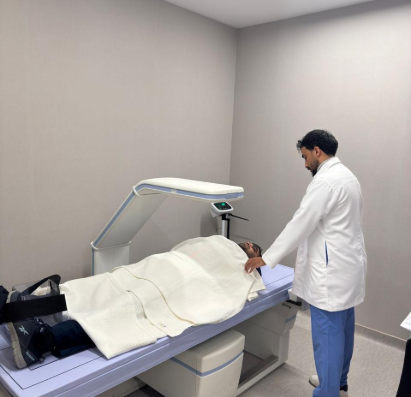
A Bone Mineral Density (BMD) and Body Composition Scan uses DXA (dual-energy X-ray absorptiometry) technology to measure bone strength and analyze body composition in a quick, painless, and non-invasive exam. The scan typically takes 5–15 minutes and uses a very low dose of X-ray radiation. This exam is particularly valuable for patients undergoing treatments that may affect bone health or body composition, including certain cancer therapies.

At OncoClinic, BMD and body composition scans are performed using carefully calibrated equipment and protocols aligned with internationally recognized diagnostic standards. This ensures results that are accurate, reliable, and clinically meaningful. These scans help our team detect early bone loss, monitor changes in muscle and fat composition, and guide supportive care strategies such as nutrition planning, physical rehabilitation, and long-term health monitoring.

What it visualizes best

  • Assessing bone strength and diagnosing osteoporosis or low bone mass
  • Early detection of bone loss before fractures occur
  • Monitoring bone health over time, especially in patients at increased risk
  • Evaluating body composition, including fat mass and lean (muscle) mass
  • Supporting personalized care plans for nutrition, exercise, and rehabilitation
shift-img

Benefits

  • Early detection of bone loss, helping prevent fractures and complications
  • Guides preventive care, including decisions about calcium, vitamin D, lifestyle changes, or medication
  • Especially helpful for patients on therapies that affect bone health, such as hormone treatments
  • Accurate assessment of fat and lean mass, supporting tailored nutrition and rehabilitation plans
  • Fast, non-invasive, and comfortable exam

Limitations

  • Provides information on bone density and body composition, but not detailed images of bones or organs
  • Does not replace other imaging exams when structural bone abnormalities or fractures are suspected
  • Results must be interpreted in the context of age, sex, medical history, and clinical risk factors

Side effect profile

  • Uses minimal radiation exposure, making it safe for repeat monitoring when clinically indicated
  • The procedure is painless and non-invasive
  • No recovery time is required—patients can return to normal activities immediately