Popular Searches
- Cancer Types
- Treatment
Popular Searches
- Cancer Types
- Treatment

Popular Searches
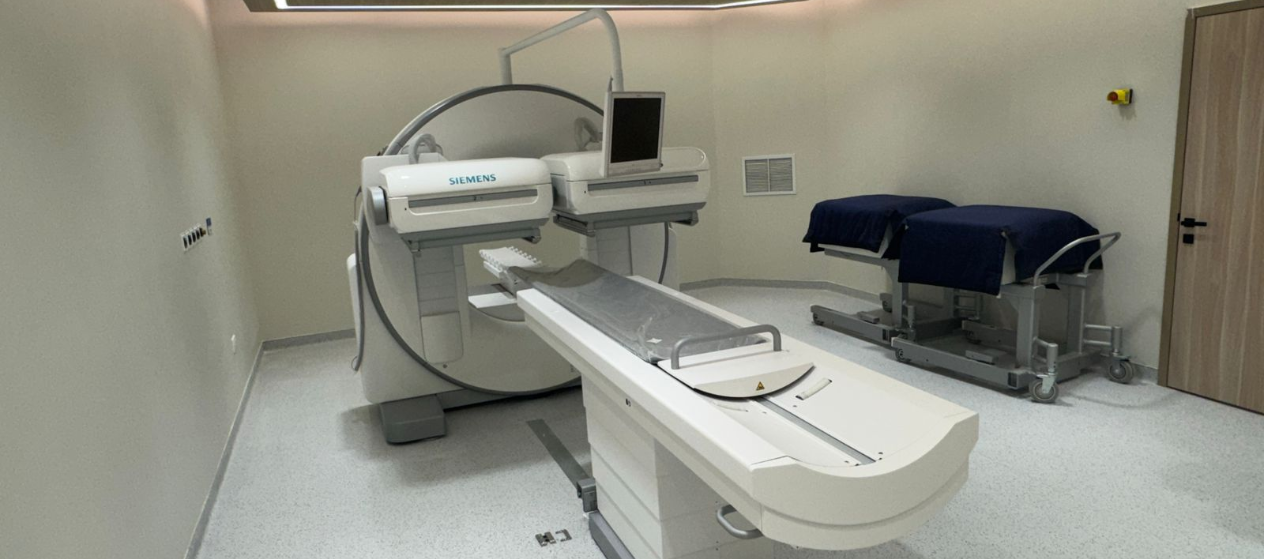
Visualizing organ function in oncological and non-oncological care.
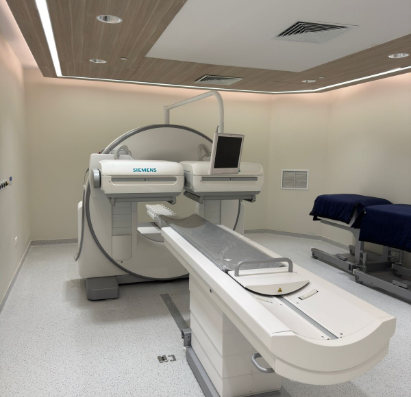
A Gamma Camera is a nuclear medicine imaging device that shows how organs and tissues are functioning, rather than only how they look. Before the scan, a small amount of a safe radioactive tracer is administered, usually by injection into the bloodstream. This tracer travels naturally to specific organs or tissues, and the gamma camera detects the signals it emits to create detailed functional images.
At OncoClinic, gamma camera imaging supports both oncological and non-oncological care, helping our specialists identify functional changes that may not yet be visible on conventional imaging such as CT or MRI. This type of imaging is especially valuable when understanding how an organ or tumor is behaving is essential for diagnosis, treatment planning, or follow-up. All gamma camera studies are performed using advanced technology and protocols aligned with international standards, ensuring safe, accurate, and reliable diagnostic information.
Bone scan: Shows changes in the bones and helps detect cancer that may have spread, giving doctors a clear view of your bone health.
The application includes:
Neuroendocrine tumor imaging (Octreoscan): Helps locate and stage neuroendocrine tumors by targeting specific receptors on tumor cells
MIBG imaging: Helps locate and assess the spread of certain cancers, allowing doctors to plan the best treatment
Lymphatic imaging: Evaluates lymphatic drainage and disease spread
Organ function assessment: Evaluates organs like the thyroid or kidneys when affected by cancer or cancer treatment
The tracer use depends on the organ being studied and the clinical question. All tracers are safe, used in very small amounts, and naturally eliminated from the body.
Technetium (Tc-99m): Most commonly used tracer in nuclear medicine, applied in bone scans, heart scans, kidney studies, and thyroid imaging due to its excellent safety profile and rapid clearance from the body.
Bone scan tracer (Technetium-based): It highlights areas of increased bone activity and is commonly used to detect bone metastases or other bone conditions.
Heart scan tracers (Technetium-based): They are used in cardiac scintigraphy to evaluate blood flow and function of the heart muscle.
Radioiodine (Iodine-123 or Iodine-131):
Octreoscan tracer: It detects neuroendocrine tumors by binding to specific receptors on tumor cells, helping identify disease location and extent.
MIBG tracer: It is used to image tumors such as neuroblastoma, pheochromocytoma, and paraganglioma.
All tracers are administered in carefully controlled doses and are typically cleared from the body within 24 hours, although radioiodine-based tracers may take slightly longer.