Logo

X-ray

Fast and effective imaging for detecting abnormalities.

contact-img

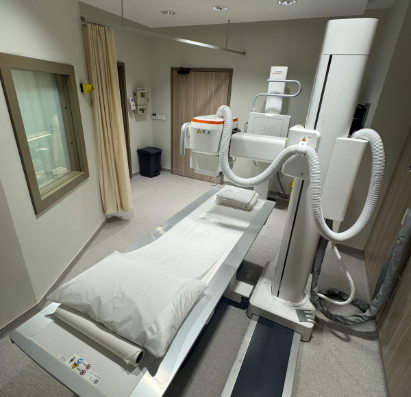
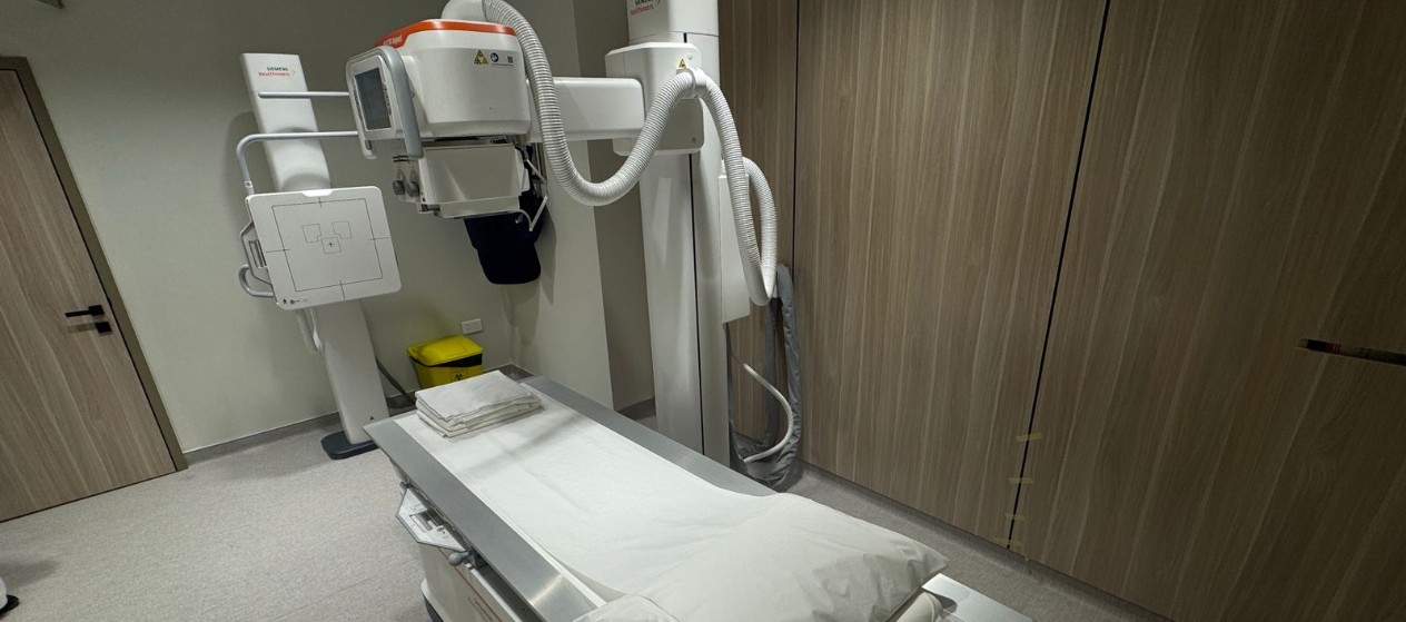
X-ray imaging is a quick, non-invasive diagnostic test that uses a small amount of radiation to create images of the body—particularly well suited for evaluating bones and lungs. It is one of the most widely used imaging tools in everyday medical care and often serves as a first-line exam to identify abnormalities or guide further testing.

At OncoClinic, we use modern X-ray equipment that follows internationally recognized quality and safety standards, ensuring clear images and timely results. X-ray imaging helps our medical team detect findings such as lung nodules, bone lesions, fractures, or other structural changes, supporting early diagnosis, treatment planning, and ongoing monitoring when appropriate.

What it visualizes best


  • Evaluation of bones: Fractures, bone lesions, and signs of cancer spread
  • Chest imaging: Lungs, ribs, and heart silhouette, including detection of nodules or fluid
  • Initial assessment: Preliminary review to identify symptoms that may require further imaging
  • Ongoing care: Follow-up and monitoring of known conditions over time
shift-img

Benefits


  • Fast and efficient imaging, with results available quickly
  • Useful first-step exam for identifying potential abnormalities
  • Low radiation dose, carefully controlled for patient safety
  • Painless, non-invasive procedure with no recovery time required

Limitations


  • Limited soft-tissue detail compared to CT or MRI
  • Provides two-dimensional images, which may not fully define the extent of disease
  • Uses ionizing radiation, though at low levels
  • Abnormal findings may require additional imaging (such as CT, MRI, or ultrasound) for further evaluation

Side effect profile


  • Uses a low dose of radiation, considered safe for most patients when clinically indicated
  • The procedure is quick and painless
  • No recovery time is required—patients can return to normal activities immediately